Related products
-
-
-
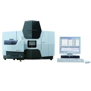
Analytical, Optical Emission Spectrometers
Optical Emission Spectrometers
There is no AI review summary. -
-
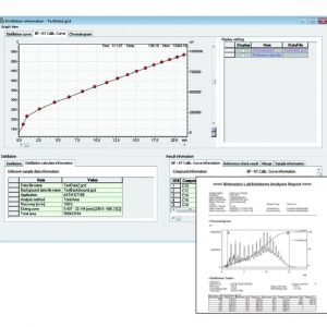
Analytical, X-ray and Surface Analysis Systems
X-ray Fluorescence Spectrometers
There is no AI review summary. -
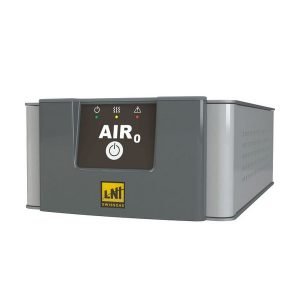
Analytical, Life Science Systems
Cell Culture Media Analysis Platform
There is no AI review summary. -
-
-
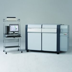
Analytical, Mass Spectrometry System
Liquid Chromatograph-Mass Spectrometers
There is no AI review summary. -
Analytical, Material Testing and Non-Destructive Inspection Systems
Universal Testing Machines
There is no AI review summary.